

Content
Research Focus
The aim of the Neuronal Core Facility is to provide state-of-the-art techniques in culturing cells from nervous tissues. Functional analysis of the cultured primary neurons has become a crucial research tool in understanding physiological and pathological changes in the central nervous system (CNS).
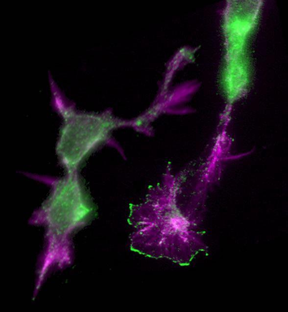
Figure 1: Immunofluorescent image of cultured cerebellar granule neurons co-stained with rhodamine-phalloidin (magenta) to visualize actin cytoskeleton and an antibody against actin regulatory protein WAVE (green).
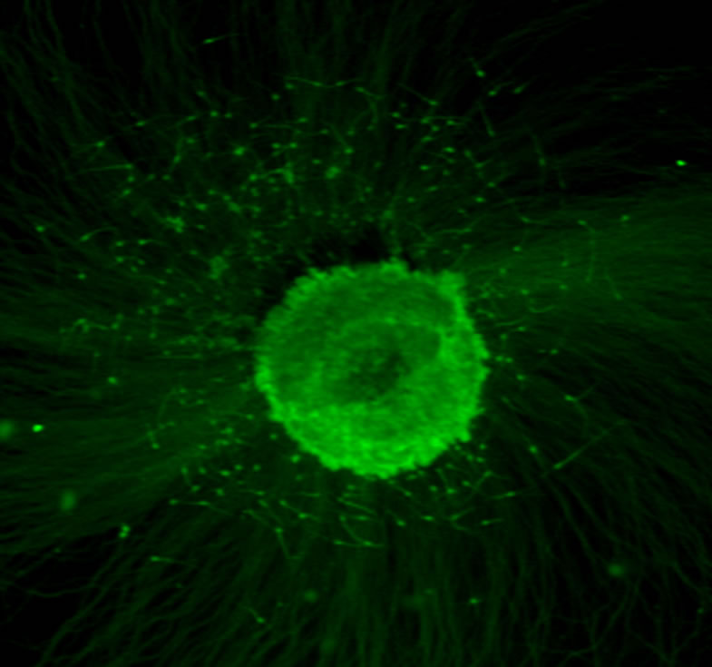
Figure 2: Immunofluorescent image of a cultured cerebellar explant immunostained with an antibody against Tuj1 to visualize migrating neurons.
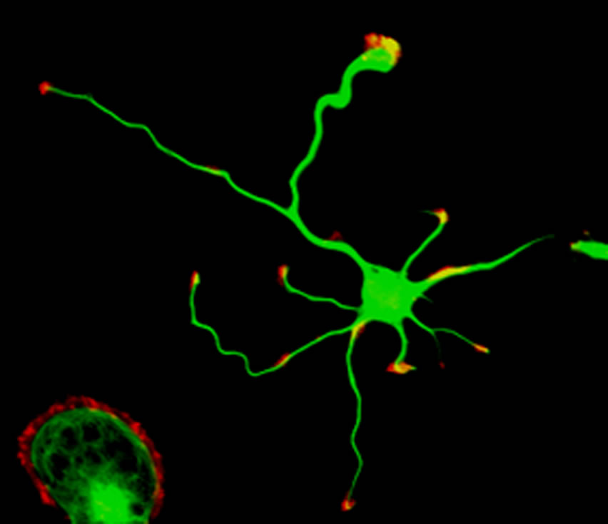
Figure 3: Immunofluorescent image of cultured hippocampal neurons co-stained with rhodamine-phalloidin (red) to visualize actin cytoskeleton and an antibody against neuronal marker Tuj1 (green).
Our priority is to facilitate research projects aimed at understanding cellular and molecular mechanisms of neurodegenerative diseases, in particular focusing at Alzheimer, Parkinson and Frontotemporal lobar degeneration (FTLD). We offer training and assistance in culturing various nervous cells such as dissociated hippocampal, cortical, cerebellar or motor neurons as well as primary astrocytes or microglia. The advantage of these primary cellular systems is that particular nerve cells are isolated and cultured under defined conditions.
Neuronal Core facility also provides training and assistance in the real-time video-microscopy of cultured neurons. This technique is beneficial in analyzing neuronal dynamics and axonal transport which, when perturbed, may contribute to neurodegeneration. Additionally, we provide expertise in explant and organotypic slice cultures in which nervous cells stay interconnected and where parts of their physiological neuronal networks remain intact.
Furthermore, we aim at improving available tools and techniques to efficiently transfect primary neurons. Currently, this is a limiting step in manipulating gene expression. In our studies, we would like to combine the functional analysis of the cultured neurons with the phenotypic analysis of the loss- or gain-of-function transgenic animal models. Both systems are valuable for understanding the physiological function of genes involved in neurodegenerative diseases. This basic understanding is also crucial for the development of disease modifying drugs. Cultured neurons offer a potential for testing the effects of these molecules on neuronal development and neurodegeneration.
Service

