

Content
Research Focus
Parkinson's disease (PD) is the second most abundant neurodegenerative disease. The disease is associated with the degeneration of dopaminergic neurons in the substantia nigra pars compacta as well as other areas of the brain. Neuropathologically PD is characterized by the accumulation of cytoplasmic deposits (Lewy bodies and Lewy neurites), which are composed of aggregating α-synuclein. It is currently unclear whether insoluble deposits or rather soluble neurotoxic oligomers contribute to disease manifestation and progression. Until recently PD was believed to be exclusively a sporadic disease associated with aging and environmental stress.
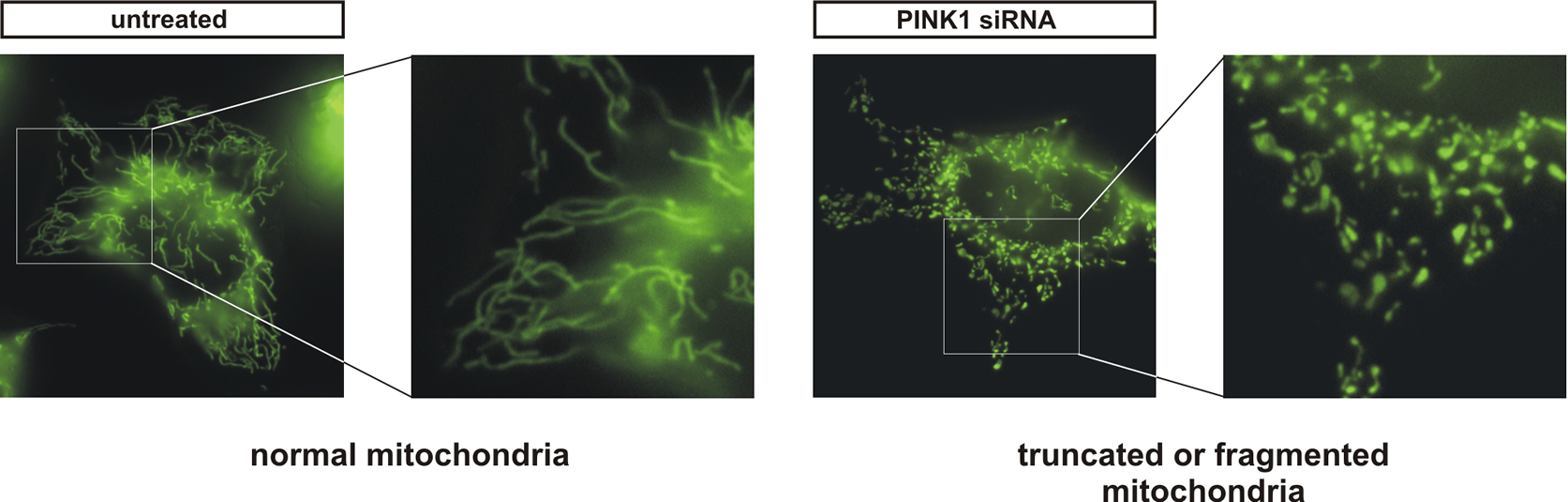
Figure 1: PINK1-deficient human cells show morphological abnormalities of mitochondria.
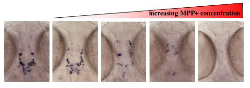
Figure 2: MPP+ treatment leads to a dose dependent loss of dopaminergic neurons in zebrafish. Dopaminergic neurons are detected by in situ hybridization using tyrosine hydroxylase as a marker.
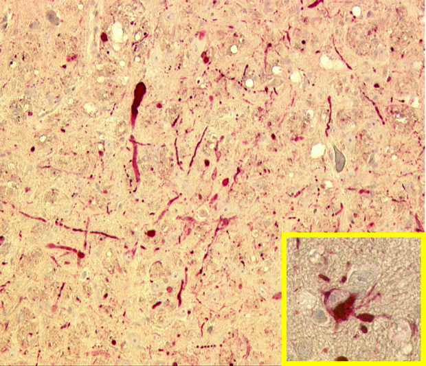
Figure 3: Pathological deposition of α-syunclein in transgenic mice expressing the A30P mutation (Neumann et al. (2002) J Clin Invest. 110(10): 1429–1439).
However, there is now very strong evidence for genetic inheritance in a small number of familial cases. Autosomal dominant mutations were so far observed in the genes encoding α-synuclein and LRRK2/dadarin. Autosomal recessive mutants associated with juvenile parkinsonism were found in parkin, DJ1, and the mitochondrial PTEN-induced kinase 1 (PINK1). PINK1 is a putative serine/threonine kinase. We are combining biochemical techniques and cell biology (Figure 1) with zebrafish (Figure 2) and mouse models (Figure 3). Using these interdisciplinary efforts we are investigating function and dysfunction of PINK1 and other PD associated genes.