

TDP-43 and FUS: a nuclear affair
Dormann D, Haass C.
21.06.2011
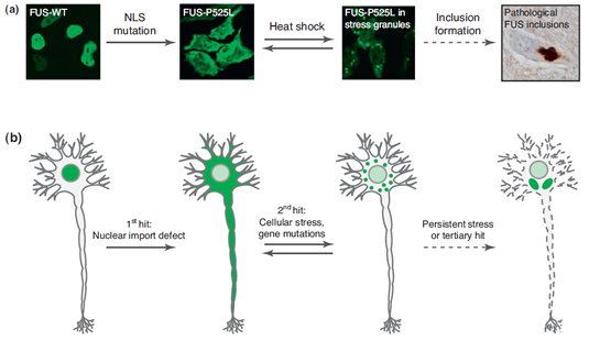
One hit is not enough: multiple-hit model of TAR DNA binding protein 43 (TDP-43)/Fused-In-Sarcoma (FUS) pathogenesis. (a) Immunocytochemistry of hemagglutinine (HA)-tagged human FUS constructs transiently transfected into HeLa cells [33]. The wild type (WT) protein is localized in the nucleus, whereas a point mutation within the proline-tyrosine nuclear localization signal (PY-NLS) of FUS (FUS-P525L) leads to cytosolic mislocalization without apparent aggregation. Upon heat shock, cytosolic FUS is recruited into stress granules [33,70], which may give rise to the large pathological FUS inclusions observed in the cytosol of ALS-FUS patients. (b) General model showing how multiple hits may lead to the formation of TDP-43 or FUS pathology. The first hit is provided by any nuclear import defect that causes a diffuse cytoplasmic localization of TDP-43 or FUS, respectively. Such defects can be genetically encoded mutations within an NLS or NES, for example the PY-NLS of FUS, or can arise sporadically during aging [e.g. by reduced expression of nuclear transport factors or oxidative damage of nucleoporins/nuclear pore complexes (NPCs)]. Moreover, post-translational modifications, such as phosphorylation, arginine methylation and proteolytic cleavage events that remove the NLS might play a role as well. A second hit that elicits cellular stress and triggers the recruitment of TDP-43 or FUS into stress granules is then required to initiate TDP-43 or FUS deposition. Such stressors might include environmental toxins or mutations in neuroprotective/stress protective genes, such as GRN or VCP. Because stress granule formation is a reversible process, this step can potentially be reversed upon release of stress or by upregulation of protective factors (e.g. chaperones or neuronal growth factors, such as Progranulin). However, persistent cellular stress or tertiary hits (e.g. genetic risk factors) may lead to the persistence of TDP-43 or FUS-containing stress granules and their conversion into large pathological TDP-43 or FUS inclusions.
ABSTRACT
Misfolded TAR DNA binding protein 43 (TDP-43) and Fused-In-Sarcoma (FUS) protein have recently been identified as pathological hallmarks of the neurodegenerative disorders amyotrophic lateral sclerosis (ALS) and frontotemporal lobar degeneration (FTLD) characterized by the presence of ubiquitin-positive inclusions (FTLD-U). Although TDP-43 and FUS are normally located predominantly in the nucleus, pathological TDP-43 and FUS inclusions are mostly found in the cytosol. Cytosolic deposition is paralleled by a striking nuclear depletion of either protein. Based on a number of recent findings, we postulate that defects in nuclear import are an important step towards TDP-43 and FUS dysfunction. Failure of nuclear transport can arise from mutations within a nuclear localization signal or from age-related decline of nuclear import mechanisms. We propose that nuclear import defects in combination with additional hits, for example cellular stress and genetic risk factors, may be a central underlying cause of ALS and FTLD-U pathology.

